Bioanalytics – a variety of methods are available for analyzing different test systems. Whether it is the quantification of single molecules, the measurement of gene activity, the localization of a protein, the determination of the mechanical properties of materials or the imaging of entire tissues, the development of new measurement methods enables the discovery of new parameters for validating, observing, and analyzing test systems while reducing the amount of starting material required for analysis. Both aspects facilitate a reduction in animal experiments by allowing in vitro models to be better studied and necessary animal experiments to be more effectively analyzed.
An important classification of measurement methods is the distinction between invasive and non-invasive applications. Non-invasive analysis methods allow for continuous measurements on the model without destroying or altering it as few as possible during data collection. In recent years, various non-invasive measurement methods have been established for different in vitro test systems, including impedance spectroscopy for measuring transepithelial electrical resistance (TEER), transport studies for investigating the barrier integrity and molecular transport of epithelial test systems, multi-electrode array (MEA) for deriving electrical signals, and analysis of cellular metabolites. Non-invasive imaging methods available include cell culture microscopy for 2D- and 3D-cultures, live-cell confocal imaging, optical coherence tomography (OCT), as well as various optical methods for in vivo systems (luminescence, fluorescence, X-ray).
Continuous measurement methods are supplemented by a variety of endpoint analyses that are associated with the destruction or strong alteration of the model. Besides classical methods such as RT-qPCR for gene expression analysis, histological staining, transmitted light, fluorescence, and electron microscopy, western blot for protein quantification, and plate rheometry to measure mechanical properties, new methods such as single-cell RNA sequencing, mass spectrometry-based proteomics, as well as new imaging techniques (light-sheet microscopy, STED, STORM) are also used for measuring endpoint analysis. In addition to developing new methods, the combination of different methods provides new approach possibilities. For example, the combination of visual imaging techniques with modern RNA sequencing methods termed spatial transcriptomics enables a spatially and histologically-resolved analysis at dimensions close to the single cell level.

© Fraunhofer ISC
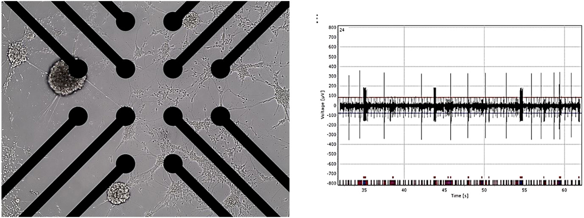
Image of a microelectrode, used to stimulate electrically active cells, such as neurons, or to record their action potentials. The right image depicts the temporally resolved electrical activity of a neuron.
